Pneumonia Indicators on X-Ray and When to Be Concerned
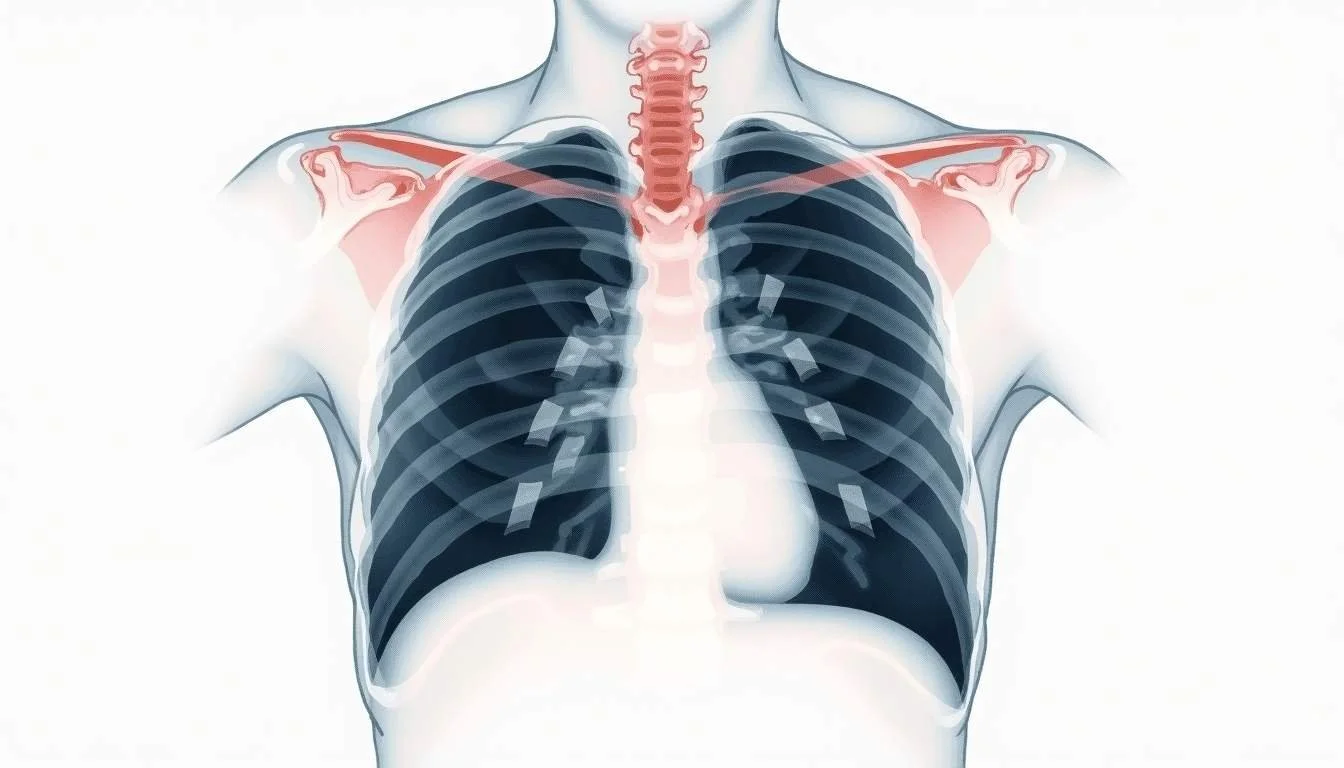
A sudden onset of breathing difficulty can send anyone into a tailspin, especially when pneumonia is a potential culprit. Chest X-rays are crucial tools for detecting pneumonia, using images that capture the internal state of the lungs. A closer look at these images provides significant insights that can inform treatment decisions. Recent studies show a variety of patterns that radiologists rely on to assess severity, including the presence of lung consolidations and other abnormalities. A notable study highlighted that over 15% of deaths among children under age five worldwide are associated with pneumonia, underscoring just how critical early detection is.
Despite advances in diagnostic technology, many cases still pose challenges for the accurate identification of severe pneumonia. With new deep learning models emerging, traditional methods are being supplemented by artificial intelligence that can provide rapid, precise evaluations. The initialization of these technologies alongside conventional methods is paving the way for improved patient outcomes and more targeted therapeutic approaches.
Recognizing the Signs on a Chest X-Ray
The human lung, when affected by pneumonia, often exhibits areas of consolidation, dense spots that indicate the presence of an infection. In radiological reports, these solid patches are among the most reliable signs of severe pneumonia. Consolidation areas are typically characterized by abnormal lung tissue opacity, which may also be accompanied by other findings such as pleural effusions or interstitial patterns.
A recent expert insight from the Journal of Cardiovascular Disease Research noted that consolidation on chest X-ray is the most sensitive indicator for severe pneumonia. This marker provides a strong negative predictive value, meaning that when consolidation is absent, severe pneumonia is less likely to be present. Identifying these markers early is key to tailoring the patient’s management plan.
Deep Learning and Automated Diagnoses
Integrating traditional radiographic techniques with deep learning is a growing area of interest among researchers. By training algorithms on large datasets of chest X-ray images, deep learning models are proving adept at differentiating between normal and severe cases of pneumonia. These models offer promise for faster and, in some cases, more accurate diagnoses than manual interpretation alone.
For instance, one study reported the development of a deep-learning-based model that successfully differentiated between normal and severe pneumonia cases. Such approaches not only enhance diagnostic precision but also potentially reduce the burden on medical professionals who already face high patient volumes. The significance of this technology is highlighted by research that demonstrates improved outcomes with earlier intervention (Detection of Pneumonia from Chest X-ray Images Using Deep Learning and Transfer Learning for Imbalanced Datasets).
These advances are significant in areas with limited access to experienced radiologists. Automated systems can offer support in rural or underserved regions, ensuring that patients receive timely care. The integration of deep learning into everyday practice marks a promising step forward in managing pneumonia and optimizing resource allocation in healthcare settings.
Comparing Imaging Findings and Patient Outcomes
Understanding Consolidation and Its Implications
Lung consolidations observed in chest X-rays are critical indicators of pneumonia severity. Research has shown a significant direct linear relationship between the presence of lung consolidations on a chest X-ray and a higher severity score. This relationship underscores why clinicians put a high emphasis on spotting such abnormalities.
When lung consolidations are identified, especially when other symptoms align with clinical findings, the diagnostic pathway tends to tilt towards aggressive treatment modalities. However, chest X-ray findings are only one part of the clinical picture. Supplemental diagnostic techniques, such as laboratory tests and patient history, are equally essential in forming a comprehensive assessment.
A study explained these dynamics in the context of COVID-19 pneumonia, stressing that consolidation patterns have a direct association with increased X-ray severity scores (Chest x-ray findings and temporal lung changes in patients with COVID-19 pneumonia). This information is particularly useful in triaging patient care during periods of high caseload in medical facilities.
Impact of Normal X-Ray Findings
Not all pneumonia patients display clear signs on an initial chest X-ray. An interesting finding from recent studies is that a significant portion of symptomatic patients may have a normal X-ray at the time of diagnosis. For example, one research study discussed that nearly 38.8% of symptomatic COVID-19 patients presented with a normal chest X-ray when initially examined (COVID-19 pneumonia study). These findings remind healthcare providers that a normal X-ray does not necessarily exclude pneumonia, especially in early stages of disease or among certain patient demographics.
This nuance is critical when assessing patient history and considering differential diagnoses. The absence of radiographic abnormalities in the early phases of an infection requires a high index of suspicion and might necessitate further evaluation through repeat imaging or additional tests. This strategy helps to ensure that cases are not missed during initial screenings.
Unsure what your scan results really mean?
We analyze MRI, CT, PET, Ultrasound, and X-ray reports and deliver a clear, easy-to-understand summary in under 1 minute.
- Understand your results in simple language
- Easy to understand explanations
- Get a list of questions to ask your doctor
Chest X-Ray Utilization in Clinical Practice
Despite the critical role chest X-rays play in diagnosing pneumonia, their application in routine family practice is not always consistent. A notable industry report observed that less than half of patients diagnosed with pneumonia by family doctors actually underwent a chest X-ray. This gap can be partly attributed to logistical challenges, limited access to radiology services, or clinical judgments based on presenting symptoms.
In many outpatient settings, especially where resources are constrained, doctors might opt for a clinical diagnosis rather than confirmatory imaging. The absence of imaging can occasionally lead to underestimation of disease severity. Patients with subtle but progressive lung changes might be misdiagnosed or their conditions under-assessed. Such scenarios highlight the need for improved access to diagnostic imaging and training in interpreting early signs on a chest X-ray.
When to Be Concerned: Red Flags on Imaging
Recognizing when a chest X-ray finding is a cause for alarm is essential, not just for medical professionals but for anyone looking to better understand their health. Several red flags on an X-ray may suggest that the pneumonia is becoming severe. One key sign is the aforementioned lung consolidation, which, when combined with worsening clinical symptoms, should prompt immediate attention.
In addition to consolidation, patients with pneumonia might show other signs like interstitial patterns and patchy infiltrates. However, it is the degree and extent of these findings that correlate with the severity of the condition. Radiologists rely on an established scoring system to evaluate the degree of lung involvement, which then informs treatment options. This scoring system can be particularly important during times when multiple patients require rapid and precise assessment.
Particularly in patients with COVID-19, chest X-rays have played a pivotal role in assessing disease severity. The direct linear relationship between consolidation presence and a higher X-ray severity score has been noted repeatedly (COVID-19 pneumonia findings). This relationship clearly shows that as the extent of consolidation increases, the risk for adverse outcomes goes up. In practical terms, careful review of these indicators should lead to more proactive treatment plans, especially for at-risk populations.
Integrating Advanced Diagnostics into Routine Care
While traditional chest X-rays remain a cornerstone of pneumonia diagnosis, the increased use of artificial intelligence is reshaping the diagnostic process. Deep learning models not only bring consistency in image interpretation but also reduce reliance on subjective human interpretation. These advancements are transforming routine care by enabling quicker identification of severe cases and tailoring treatment measures accordingly.
The ability of a deep-learning-based model to distinguish normal from severe pneumonia cases offers an improved approach to triaging patients. By automating part of the diagnostic process, healthcare workers are provided with an extra layer of confirmation that can be especially valuable in busy medical centers. This innovation holds significant promise for improving patient outcomes by ensuring that those with severe presentations receive timely intervention.
Adoption of such advanced diagnostics is still evolving, but the science behind it is robust. When conventional evaluations are combined with deep learning, the resulting synergy can streamline decisions in both hospital and community settings. Such integration is increasingly necessary in a healthcare landscape where rapid, informed decision-making has become a vital component of patient care.
Practical Considerations for Clinicians and Patients
Improving Diagnostic Accuracy
For clinicians, improving diagnostic accuracy means a combination of careful imaging interpretation, advanced technology, and clinical judgment. While the presence of consolidation is a prominent indicator of pneumonia severity, the absence of these changes on an X-ray does not guarantee that a patient is free from the disease. This gap reinforces the need for periodic reassessments, especially in patients whose clinical symptoms persist or worsen.
Clinicians are encouraged to consider additional diagnostic tests if initial imaging results are inconclusive. Laboratory tests, including inflammatory markers and cultures, can supplement imaging findings. By correlating imaging studies with clinical presentations and lab values, the overall diagnostic picture becomes clearer and more actionable. This comprehensive approach ensures that patients receive accurate diagnoses and appropriate treatment.
The Role of Follow-Up and Reassessment
Once pneumonia is suspected or diagnosed, follow-up imaging often becomes necessary to monitor progress or resolution of the infection. A repeat chest X-ray can help determine whether the patient’s condition is improving or if further treatment adjustments are needed. Even mild findings can progress if left unchecked; thus, periodic reassessment is key.
In some cases, patients who initially show clear signs of pneumonia might display only subtle abnormalities during follow-up. This phenomenon is a reminder that changes in lung appearance often have a temporal evolution. Early consolidation may give way to more diffuse patterns or even a return to near-normal imaging in cases of mild-to-moderate disease. Understanding these patterns helps clinicians make more informed decisions about the duration of therapy and the need for additional intervention.
The Broader Impact of Imaging on Pneumonia Management
The evolution of chest X-ray technology and its integration with modern artificial intelligence tools have a broader impact on public health. Not only does it improve individual case management, but it also supports better epidemiological tracking of pneumonia outbreaks. Careful documentation and analysis of chest X-ray findings across populations help identify trends in disease severity and treatment effectiveness.
For example, studies have shown that chest X-rays play a significant role in assessing the progression of pneumonia in various populations, from children to the elderly. This long-term data contributes to better health strategies and can guide resource allocation during epidemic periods or seasonal surges. Improved diagnostic protocols help reduce misdiagnoses and enhance the overall quality of care delivered within communities.
Linking diagnostic improvements directly to public health outcomes creates a cycle of benefit that extends well beyond the immediate clinical setting. Innovations in radiographic technology and analysis also empower healthcare workers with reliable data, leading to refined treatment protocols and, ultimately, improved patient survival rates.
Looking Ahead: Innovations and Future Directions
As medical imaging evolves, the potential for even more accurate detection of pneumonia grows. Future directions likely include further refinement of deep learning models and their integration with other advanced imaging modalities. Emerging research continues to push the boundaries of what is possible, offering hope for even faster and more reliable diagnostic methods.
The combined efforts of radiologists, computer scientists, and clinicians continue to drive forward our understanding of pneumonia and other respiratory diseases. These collaborations are not only advancing imaging technology but also bridging gaps in access to care, especially in remote and underserved areas. Findings from recent studies, such as those linking lung consolidation with disease severity (chest X-ray research), provide a foundation for these ongoing improvements.
Looking ahead, the landscape of pneumonia management is likely to see an even greater emphasis on personalized medicine, where imaging, laboratory data, and patient-specific factors combine to drive treatment. New algorithms may soon offer predictive insights, guiding both diagnostic and therapeutic decisions with unprecedented precision.
Final Thoughts: Empowering Decisions Through Imaging
Chest X-rays remain a critical component in diagnosing pneumonia, offering a window into the unspoken signs of lung infections. As the medical field adopts newer, more automated approaches alongside traditional techniques, the accuracy and timeliness of pneumonia diagnoses are only set to improve. Imaging not only aids in determining the severity of the infection but also supports a more tailored approach to treatment.
Recognizing key radiographic patterns such as lung consolidation is essential. When these signs are detected quickly, the subsequent management can be aggressive enough to save lives, especially in vulnerable populations. As highlighted in the initial study linking pneumonia to significant mortality rates among children (Identifying pneumonia in chest X-rays), timely and accurate diagnosis is paramount.
For both clinicians and patients, understanding the nuances of chest X-ray findings translates into more informed healthcare decisions. With advancements in technology and a clearer grasp of how imaging correlates with clinical severity, the fight against pneumonia becomes increasingly data-driven and precise. It is an approach that promises not only better individual outcomes but also a significant step forward in public health management.
This evolving dialogue between technology and clinical practice ensures that pneumonia management will continue to improve. Each breakthrough in radiological imaging brings with it the potential for better detection, earlier treatment, and ultimately, improved survival rates. The integration of these advancements into everyday practice is a testament to the continual striving for excellence in patient care.
Waiting for answers? You don’t have to.
Upload your MRI, CT, PET, Ultrasound, or X-ray report and receive a clear explanation in < 1 minute.
Simplify Your Chest X-Ray Understanding with Read My MRI
If you're grappling with the complexities of a chest X-ray report and suspect pneumonia, Read My MRI is here to help. Our AI-driven platform can translate your medical imaging reports into straightforward, jargon-free summaries. Gain peace of mind and clarity on your health concerns by using Read My MRI to get a clear, concise understanding of your imaging results. Get Your AI MRI Report Now! and take the first step towards empowered health decisions.