Chest X-Ray Reveals Spot on Lung: Cancer or Something Else?
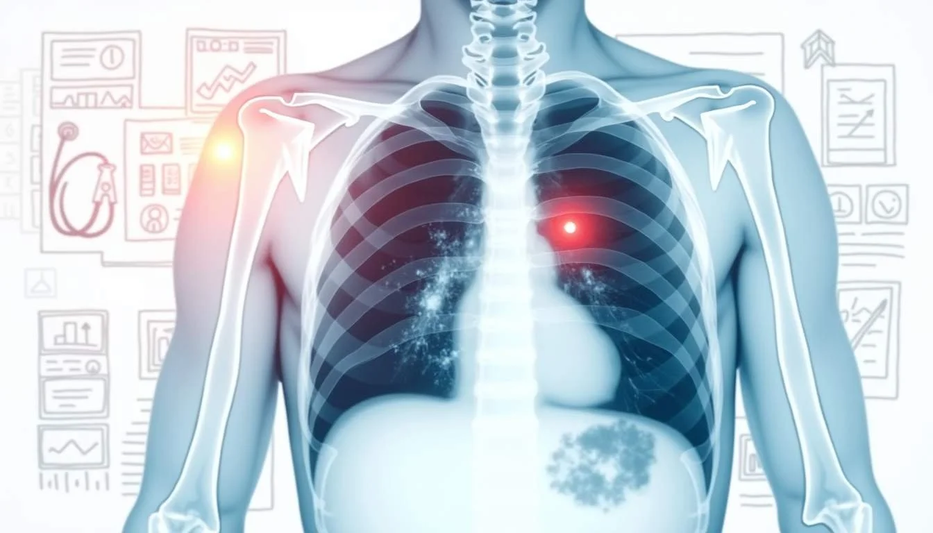
A routine chest X-ray can sometimes uncover an unexpected spot on the lung, raising immediate concerns. Questions emerge quickly-does this indicate cancer, or could it be a benign finding? With modern technologies and evolving research insights, doctors are better able to guide patients through these uncertain moments.
Recent advances, particularly in artificial intelligence, are playing a big role in clarifying these situations. A study published by the Radiological Society of North America (RSNA) showcased how AI-based software significantly improved the detection of lung nodules on chest radiographs.
Understanding Lung Nodules
Lung nodules are small masses of tissue that appear on imaging tests. They may result from infections, benign growths, or even early manifestations of cancer. Before jumping to conclusions, it's crucial to understand these nodules are quite common, even among those who have never smoked. For example, research in Diagnostic Imaging revealed that over 40% of non-smokers have at least one lung nodule, with around 11.1% having clinically relevant nodules (Diagnostic Imaging).
This statistic alone provides a framework to understand why a single nodule does not immediately signal cancer. Clinicians analyze a host of factors, including the nodule's size, shape, and location, as well as the patient's clinical history. The mere presence of a nodule triggers a series of diagnostic protocols to determine its nature.
Over time, technological advancements and refined imaging techniques have helped differentiate harmless nodules from those requiring further inspection or treatment.
The Role of Advanced Imaging and AI
Enhancing Diagnostic Accuracy
Imaging technology has evolved beyond simply producing pictures. Modern chest X-rays and CT scans are high-resolution tools that offer detailed views of lung anatomy. Such precision is especially valuable when interpreting subtle abnormalities.
Recent studies highlighted by the RSNA have shown that software driven by artificial intelligence can enhance the detection of lung nodules considerably. In one study, around 1% of patients, specifically 152 out of 14,563, had unexpected lung nodules picked up by AI on chest radiographs (PubMed).
This technology acts as a second pair of eyes, complementing the radiologist’s expertise. By flagging potential issues more reliably, AI helps reduce human error and accelerates diagnostic decisions.
How AI Improves Detection
Traditional methods of reading radiographs have been enhanced by AI algorithms that learn from vast libraries of images. These algorithms are trained to recognize patterns that might be missed by the naked eye. The introduction of these tools in everyday diagnostics has been particularly beneficial in busy clinical settings where high patient volumes might otherwise lead to oversights.
AI-based software not only increases detection accuracy but also standardizes the evaluation of chest X-rays, reducing subjective differences among radiologists. As lung nodules can be an early sign of cancer, the ability to catch them at an early stage is a game-changer in the management of lung health.
The integration of advanced imaging technologies with AI drives improvements across the board, offering hope and reassurance to patients who may be facing uncertain diagnoses.
Distinguishing Between Benign and Malignant Nodules
Given that lung nodules are not uncommon, the challenge for physicians is determining which nodules warrant further attention. While some nodules pose little risk, others might signal the onset of something more serious, like lung cancer. This difference underscores the importance of a comprehensive diagnostic approach.
Radiologists use a combination of imaging characteristics and patient history to make these distinctions. Nodules are most often monitored over time with serial imaging to assess changes in size or appearance. For example, a nodule that remains stable over several imaging sessions is less likely to be malignant. This observation-based method is crucial because lung cancer often needs to be caught early for effective treatment.
Research has shown that subtle changes detected by advanced imaging tools might indicate a progression that warrants biopsy or further testing. Hence, the integration of precise tools and vigilant monitoring strategies is central to the approach.
Unsure what your scan results really mean?
We analyze MRI, CT, PET, Ultrasound, and X-ray reports and deliver a clear, easy-to-understand summary in under 1 minute.
- Understand your results in simple language
- Easy to understand explanations
- Get a list of questions to ask your doctor
Evaluating the Broader Impact of AI in Chest Imaging
The ripple effect of incorporating AI into chest imaging is far-reaching. Hospitals and healthcare centers across the United States perform millions of chest X-rays and CT scans annually. UCLA Health reports that over 70 million chest X-rays and 80 million CT scans are conducted each year, with nearly 50% of these scans revealing lung nodules (UCLA Health).
The sheer volume of these scans underscores the need for efficient, accurate methods of analysis. By incorporating AI, radiologists can quickly pinpoint areas of concern, thereby expediting the diagnostic process. This efficiency is critical in scenarios where early detection of lung cancer can be life-saving.
Improved diagnostic performance not only enhances individual patient care but also optimizes healthcare resources. The ability to quickly rule in or out the need for further invasive tests can reduce unnecessary procedures, contributing to overall better patient management.
Implications for Patient Care and Future Trends
The evolving landscape of imaging diagnostics has significant implications for patient care. When lung nodules are detected, the immediate reaction is often fear. However, the increasing accuracy of imaging and the help provided by AI reduces the uncertainty, allowing for timely and appropriate management.
Patients with a detected lung nodule may undergo follow-up scans, additional tests, or a biopsy to determine its nature. These steps are part of a broader strategy to ensure that, if it is cancer, it is caught as early as possible, making treatment more feasible. As one expert insight noted from the RSNA, AI's role in accurately interpreting chest radiography could significantly bolster early identification of chest diseases.
With advances in technology and robust research supporting new diagnostic strategies, future trends point toward a more integrated and technology-driven approach in evaluating chest imaging. This integration symbolizes a future where precision medicine is the norm, minimizing the risk of misdiagnosis and enabling tailored treatment plans for each patient.
The convergence of technological innovation and clinical expertise is redefining patient journeys. Even when a chest X-ray reveals a suspicious spot, the streamlined process of follow-up and diagnosis allows for a measured response, where anxiety is replaced by clarity and direction.
The Psychological Impact on Patients
The discovery of a lung nodule on an X-ray naturally sparks fear and anxiety. For many individuals, the possibility of lung cancer evokes intense emotional responses. Healthcare providers must approach these situations with sensitivity, ensuring patients are well-informed.
Clear communication regarding the prevalence of lung nodules can be comforting. When patients understand that lung nodules are common, even among non-smokers, it can help contextualize their personal situation. For instance, research detailed by Diagnostic Imaging shows that many people without a smoking history have lung nodules, most of which are non-cancerous.
Medical professionals are encouraged to discuss the nature of these findings thoroughly. They offer guidance on follow-up plans and, when needed, stress the benefits of modern diagnostic tools such as AI to alleviate the burden of uncertainty. This transparent dialogue helps patients engage with their health in a more proactive and less fearful manner.
Navigating Next Steps After a Nodule Is Found
Once a lung nodule has been identified, several treatment options may be recommended. The decision-making process typically begins with a more detailed imaging scan, such as a CT scan, to better assess the nodule's features. Follow-up imaging allows clinicians to monitor for any radiological changes over time. A change in size or appearance might suggest the need for an invasive procedure, like a biopsy, to definitively rule out cancer.
The management strategy is deeply personalized. For many, the initial identification of a nodule will lead to a routine follow-up schedule rather than immediate aggressive intervention. The use of AI in this process is proving valuable once again. By consistently evaluating chest X-rays, AI supports a more rapid and accurate decision-making process, ensuring that those who need more urgent care receive it promptly.
This cautious approach, balancing vigilance with the understanding that most nodules are benign, helps avoid unnecessary stress and invasive procedures. Health systems, by leveraging high-volume imaging and advanced analytical techniques, can more effectively differentiate nodules that require intensive follow-up from those that can be safely observed.
The Broader Picture: Public Health and Screening Initiatives
The impact of advancing imaging technology extends beyond individual diagnoses; it plays a crucial role in public health. Regular screening, especially for high-risk groups, is essential for early detection of lung cancer. With the capability of AI to enhance assessments, routine imaging tests become even more valuable tools for early intervention.
Healthcare providers now have access to robust data showing that early detection through advanced imaging can lead to better outcomes. Current practices often rely on a layered approach-using both chest X-rays and CT scans-to ensure no potential abnormality is overlooked. The integration of AI further refines this process, offering both breadth and precision in the analysis.
As technology improves and more data become available, public health initiatives can adopt increasingly tailored screening protocols. This potential shift in screening methodology might lead to reduced mortality from lung cancer by catching it at a stage when treatment is most effective. The focus is not solely on detection, but also on the rapid and appropriate follow-up, ensuring that the window for early intervention does not close.
Putting It All Together
Deciding whether a spot on a lung is cancerous or benign is a multidimensional process. It involves a combination of advanced imaging techniques, a careful review of medical history, and the increasingly pivotal role of artificial intelligence. With each advancement, the healthcare community steps closer to providing patients with more accurate diagnoses and shorter wait times.
Statistics and research have played a significant role in shaping this nuanced understanding of lung nodules. Consider that even among non-smokers, where one might expect lower risk, nearly half of these individuals may display at least one lung nodule (Diagnostic Imaging). This insight drives home the importance of not jumping to conclusions and underscores the need for thorough evaluations.
The evolution of imaging diagnostics from simple X-rays to AI-enhanced evaluations illustrates the remarkable strides taken in lung health. The presence of AI in diagnostic pathways has been validated by multiple studies, showing improvements in accuracy that can have a direct impact on patient outcomes. With a careful balance between technology and clinical judgment, the approach to managing lung nodules continues to evolve, offering hope to many who might otherwise face uncertainty.
Looking Ahead: The Future of Lung Nodule Detection
The future of lung nodule detection promises to be even more advanced. Research continues to push the boundaries of what is possible with imaging technology. As AI algorithms become more sophisticated, they will likely be able to provide even earlier and more accurate assessments. The potential for combining imaging data with other clinical information could lead to personalized diagnostic and treatment plans tailored exactly to each patient’s needs.
Health systems are already beginning to see the benefits of these advancements. With routine screenings enhanced by AI and systems designed to monitor changes in lung nodules over time, the outlook is optimistic. This technological progression not only supports early detection of potential cancers but also helps in reducing unnecessary interventions by clearly distinguishing between benign and malignant nodules.
Future clinical practices may see a seamless integration of patient history, imaging data, and real-time analytics-a convergence that brings personalized medicine to the forefront. As these practices become standardized, the hope is that more lives will be saved by catching potential cancers before they progress to an advanced stage.
Empowering Patients Through Knowledge
Understanding the role of imaging in detecting lung abnormalities can empower patients. Knowledge about what lung nodules are, how frequently they occur, and what the next steps involve can turn anxiety into informed decision-making. Making fact-based decisions becomes easier when patients realize that a spot on the lung is a starting point for more detailed scrutiny rather than a definitive diagnosis of cancer.
Accurate information, backed by reliable studies such as those highlighted by RSNA, reassures individuals facing a lung nodule diagnosis. Moreover, experts emphasize that the early detection provided by such technologies not only aids in the swift management of potential lung cancer but also minimizes the risk of overtreatment for benign conditions.
Awareness is a powerful tool. Educating the public about the prevalence of lung nodules and the advances in medical imaging technology fosters trust in the diagnostic process. While the discovery of an abnormal spot can be unsettling, patients can find comfort in the fact that modern medicine is well-equipped to determine its nature.
Chest X-rays continue to be an essential component of lung health assessments, serving both as an initial screening tool and as part of a comprehensive diagnostic strategy. The discovery of a lung nodule initiates a careful, multi-step evaluation process that relies on both human expertise and technological advancements. With the integration of AI technologies, the detection and monitoring of lung nodules have become more precise, ultimately contributing to improved patient outcomes.
Though the presence of a nodule can spark immediate concern, the broader picture is one of cautious optimism. Research highlights, whether it’s the robust statistics from Diagnostic Imaging or the promising study outcomes from RSNA, affirm that modern medicine is ever-improving. Such advances are instrumental in ensuring that every lung nodule is evaluated with the highest possible accuracy.
For anyone facing an uncertain diagnosis, it is reassuring to know that a rigorous, technology-driven medical process mitigates the apparent risks associated with a lung nodule. The balance of evidence, careful monitoring, and rapid follow-up makes it clear that an isolated spot is just one piece of a larger diagnostic journey-a journey where each step is designed to ensure the safest possible outcome for every patient.
Waiting for answers? You don’t have to.
Upload your MRI, CT, PET, Ultrasound, or X-ray report and receive a clear explanation in < 1 minute.
Understand Your Lung Health with Ease
If you've had a lung screening and are puzzled by the complex medical terminology in your report, Read My MRI is here to help. Our AI-powered platform simplifies your MRI, CT, PET, X-Ray, or Ultrasound reports, providing you with clear, jargon-free summaries. Gain peace of mind and a better understanding of your lung health. Get Your AI MRI Report Now!