How Do I Understand My PET Scan Report?
Getting a Positron Emission Tomography (PET) scan can feel like stepping into a world of complex medical jargon and mysterious images. When the report lands in your hands, it might look like a foreign language. But understanding your PET scan report is crucial-it helps you grasp what your doctors see and guides your next steps in health care.
With the PET scanner market growing rapidly and innovations like Siemens Healthineers’ Biograph Vision.X improving image quality and speed, these scans are becoming more accurate and accessible than ever before. This guide breaks down the essentials to help you make sense of your PET scan report with confidence. For a detailed overview of the PET market and technology trends, you can explore this market report.
What Is a PET Scan and Why Is It Used?
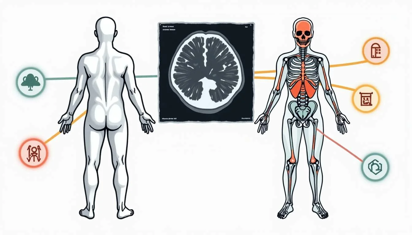
A PET scan is a type of imaging test that helps doctors see how your tissues and organs are functioning. Unlike traditional X-rays or CT scans that show structure, PET scans reveal metabolic activity by detecting radioactive tracers injected into your body. This makes PET scans especially valuable in detecting cancer, heart problems, brain disorders, and infections.
For example, in oncology, PET scans can identify cancerous cells because they consume more glucose than normal cells, lighting up on the scan. This functional insight often leads to earlier and more precise diagnoses.
Thanks to advances in technology, such as the enhanced time-of-flight capabilities introduced by Siemens Healthineers in 2023, PET scans now deliver sharper images faster, improving diagnostic accuracy and patient comfort. Learn more about these innovations here.
In addition to its applications in oncology, PET scans are also crucial in cardiology. They can assess the heart's function and blood flow, helping to identify areas of reduced blood supply that may indicate coronary artery disease. This information can guide treatment decisions, such as whether a patient may benefit from angioplasty or bypass surgery. Moreover, PET scans are increasingly being used in the evaluation of neurodegenerative diseases like Alzheimer's, where they can help visualize amyloid plaques in the brain, providing insights into the disease's progression and aiding in the development of targeted therapies.
The procedure itself is generally well-tolerated, though patients may experience some anxiety due to the injection of the radioactive tracer. However, the amount of radiation exposure is typically low, comparable to that of a standard CT scan. As a result, the benefits of obtaining critical diagnostic information often outweigh the risks. Furthermore, many facilities are now incorporating patient-friendly practices, such as providing calming environments and pre-scan education, to enhance the overall experience and alleviate any concerns patients may have about the procedure.
Unsure what your scan results really mean?
We analyze MRI, CT, PET, Ultrasound, and X-ray reports and deliver a clear, easy-to-understand summary in under 1 minute.
- Understand your results in simple language
- Easy to understand explanations
- Get a list of questions to ask your doctor
Breaking Down Your PET Scan Report
Your PET scan report usually contains several key sections: patient information, scan details, findings, impression, and recommendations. Understanding each part can demystify the report.
1. Patient Information and Scan Details
This section includes your name, date of birth, the date of the scan, and sometimes the reason for the scan. It also details the type of PET scan performed—sometimes combined with CT (PET/CT)—and the tracer used. For instance, PSMA-PET/CT is a specialized scan for prostate cancer detection, which has shown promising results with an 80% sensitivity in recent studies.
Knowing the tracer and scan type helps you understand what the images are highlighting and why. Different tracers are used for various conditions; for example, FDG (fluorodeoxyglucose) is commonly used for detecting a variety of cancers, while others like NaF (sodium fluoride) are more specific for bone metastases. This knowledge not only helps you grasp the focus of the scan but also prepares you for discussions with your healthcare provider about what the results might mean for your health journey.
2. Findings
This is the heart of the report, where radiologists describe what they see. They note areas of increased or decreased tracer uptake, size and location of any lesions, and abnormalities. Increased uptake can indicate cancer, inflammation, or infection, but it’s not always cancer—sometimes benign conditions light up too.
For example, a study on metastatic prostate cancer detection using multi-modal volumetric concept activation on PSMA-PET/CT achieved a sensitivity of 80% but also reported some false positives, emphasizing the importance of clinical correlation. Radiologists often employ advanced imaging techniques and software to enhance the accuracy of their findings. They may also compare current scans with previous ones to assess changes over time, which can be crucial in determining the progression of a disease or the effectiveness of treatment.
Understanding that findings are descriptive and need interpretation in context is key. Each patient's medical history and symptoms play a significant role in how these findings are viewed, making it essential to engage in an open dialogue with your healthcare team.
3. Impression
This section summarizes the main conclusions. It often states whether the scan is normal or if suspicious areas warrant further investigation. The impression guides your doctor’s recommendations and next steps. It is important to note that while the impression is based on the radiologist's expertise, it is still just one piece of the puzzle in your overall health assessment.
In some cases, the impression may suggest the need for additional imaging studies or consultations with specialists, depending on what the findings indicate. This proactive approach allows for a comprehensive evaluation of your condition, ensuring that no potential issues are overlooked.
4. Recommendations
Based on the findings and impression, the report may suggest follow-up scans, biopsies, or treatments. This part is crucial for planning your care. Recommendations are tailored to your individual situation and can vary significantly depending on the results of your scan. For instance, if a suspicious lesion is identified, your doctor may recommend a biopsy to obtain a definitive diagnosis.
Additionally, recommendations may also include lifestyle changes, such as dietary adjustments or exercise regimens, which can play a supportive role in your overall treatment plan. It's essential to discuss these recommendations thoroughly with your healthcare provider to understand the rationale behind each suggestion and how they fit into your broader health strategy. Engaging actively in this process can empower you to make informed decisions about your care moving forward.
How to Interpret Common Terms and Results
PET scan reports are filled with medical terms that can be confusing. Here are some common phrases and what they generally mean:
Hypermetabolic focus: An area showing higher than normal tracer uptake, possibly indicating cancer or inflammation.
Hypometabolic area: A region with less uptake, which could suggest tissue damage or necrosis.
Standardized Uptake Value (SUV): A number indicating the intensity of tracer uptake. Higher SUV values often raise suspicion but aren’t definitive alone.
False positives: Areas that appear suspicious but are benign. Studies highlight that false positives can occur, so doctors consider the whole clinical picture.
Remember, no single number or finding tells the whole story. Radiologists combine these observations with your history, symptoms, and other tests.
The Role of Double Reading and AI in Enhancing Accuracy
Reading a PET scan is complex. A recent multicenter study involving 678 patients found that systematic double reading-where two experts independently review the scan-significantly improved diagnostic accuracy. This approach reduces errors and ensures subtle findings aren’t missed.
Artificial intelligence (AI) is also transforming PET imaging. AI algorithms assist in image reconstruction, lesion detection, and classification, enhancing both speed and precision. Experts note that AI has significant potential to advance PET imaging applications, helping radiologists interpret scans more reliably.
As the PET scanner market continues to innovate, integrating AI and advanced technologies like those from Siemens Healthineers, patients benefit from more accurate and faster diagnoses. For more on AI’s impact in medical imaging, see this expert insight.
Understanding Your PET Scan in the Context of Your Health
Your PET scan report is one piece of the puzzle. It complements other tests and clinical evaluations. For example, in cancer care, PET scans help stage the disease, monitor treatment response, and detect recurrence.
In North America, where the PET market is the largest globally, valued at over USD 665 million in 2022 and expected to exceed USD 1 billion by 2031, these scans are increasingly integrated into routine oncological care. This growth reflects their proven value in improving patient outcomes.
Always discuss your report with your healthcare provider. They can explain what the findings mean for your specific situation and recommend the best course of action.
What to Ask Your Doctor After Receiving Your PET Scan Report
It’s normal to have questions once you see your PET scan report. Here are some useful questions to consider:
What do the areas of increased or decreased uptake mean in my case?
Are there any suspicious findings that require further testing?
How do these results affect my diagnosis or treatment plan?
Is a biopsy or follow-up scan necessary?
How reliable are the findings-was the scan double-read or assisted by AI?
Being proactive and informed helps you engage in your care effectively.
Looking Ahead: The Future of PET Scanning
The PET scanning field is advancing rapidly. With a projected market growth rate of over 5% annually through 2031, driven by technological innovation and AI integration, PET scans will become even more precise and accessible.
New scanners like the Biograph Vision.X are setting new standards for image quality and speed. Meanwhile, research continues to refine how PET scans detect and classify diseases, improving sensitivity and reducing false positives.
For those interested in market trends and technology developments, this industry report offers a comprehensive look at the evolving landscape.
Understanding your PET scan report empowers you to take control of your health journey. With clearer insights and ongoing advancements, these scans are invaluable tools in modern medicine.
Take Charge of Your Health with Read My MRI
Understanding your PET scan report is just the beginning of your health journey. At Read My MRI, we're dedicated to simplifying this process for you. Our AI-powered platform turns complex medical reports from MRI, CT, PET, X-Ray, or Ultrasound into easy-to-comprehend summaries. Say goodbye to the confusion of medical terms and embrace clear, jargon-free explanations. Whether you're deciphering your own health information or a healthcare provider streamlining report analysis, Read My MRI is your ally in making medical reports understandable for everyone. Ready to gain clarity on your diagnosis? Get Your AI MRI Report Now!
Waiting for answers? You don’t have to.
Upload your MRI, CT, PET, Ultrasound, or X-ray report and receive a clear explanation in < 1 minute.