MRI Shows Demyelination: Should You Panic?
The MRI report lands in the patient portal, and the eyes go straight to one terrifying phrase: “findings suggest demyelination.” Heart rate spikes, search history fills with “multiple sclerosis,” and it suddenly feels like everything has changed. Yet that single word on a scan report is only one piece of a much bigger picture, and it does not automatically equal a diagnosis, disability, or a predictable future.
Radiology language is written for other clinicians, not for patients. It tends to sound dramatic, even when the actual risk is uncertain or low. Even with multiple sclerosis (MS), which is the condition most people think of when they see “demyelination,” MRI is far from perfect. The National Multiple Sclerosis Society notes that a small but real group of people can meet clear clinical criteria for MS yet have essentially normal MRI scans, which shows how much nuance sits behind a simple report line.
What “demyelination” on MRI actually means
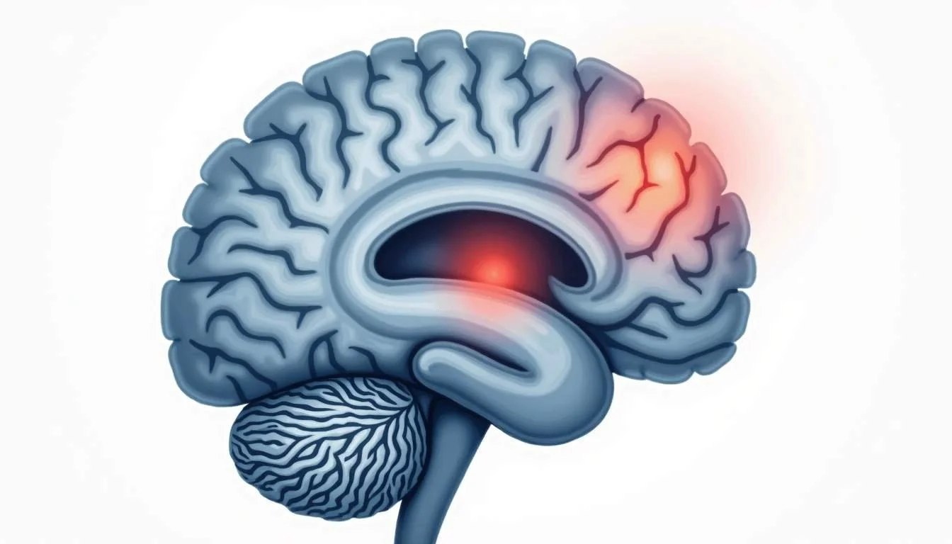
Myelin is the protective coating wrapped around nerve fibers in the brain and spinal cord. It works like insulation around electrical wiring, helping signals travel quickly and efficiently. When that coating is damaged or lost, the process is called demyelination. On MRI, radiologists do not see myelin itself; they see changes in water content, tissue structure, and other indirect signals that suggest myelin has been disrupted.
That distinction matters. An MRI showing demyelination describes a pattern that often fits MS, but it can also appear with other conditions: migraine, small-vessel disease, infections, autoimmune disorders outside the typical MS spectrum, and even severe vitamin deficiencies. Advanced MRI techniques are getting better at teasing these processes apart. Conference reports describe how specialized approaches such as magnetization transfer, diffusion imaging, and myelin mapping are revealing different aspects of tissue damage in MS and related disorders, offering a more refined view than standard scans alone can provide, as summarized in an industry overview of advanced MRI in MS.
Research looking directly at myelin density in people with MS has shown that multiple MRI parameters-such as measures from T1 and T2 imaging, magnetization transfer, and diffusion techniques-tend to track reasonably well with how much myelin is present in the tissue that was sampled. A recent analysis reported that several of these parameters show moderate to strong correlations with myelin density, suggesting that MRI is increasingly capable of capturing real biological change rather than just vague “spots” on the brain, according to a study of MRI markers and myelin density.
How reliable is MRI for detecting MS?
For MS, MRI is an essential tool-but not an infallible one. It is very good at revealing areas where the immune system has attacked myelin in the brain or spinal cord. It is less good at telling doctors exactly what caused that pattern, especially if there are only a few spots or if they do not follow classic MS rules. It is also not guaranteed to show abnormalities even when MS is definitely present clinically.
Guidance from the National Multiple Sclerosis Society notes that a small percentage of people with clinically definite MS-people whose symptoms and neurologic exams clearly fit the disease-do not show lesions on MRI at all. That reminder cuts two ways. It means a “clean” MRI does not rule MS out when everything else points toward it. It also means a scan that looks suspicious is still only part of the diagnosis and must be interpreted alongside history, symptoms, and exam findings.
Another piece of this puzzle is radiologically isolated syndrome (RIS). In RIS, brain or spinal cord MRI scans look strikingly similar to MS, yet the person has no neurological symptoms. When researchers looked across multiple MRI modalities and large groups of scanned individuals, the cumulative incidence of RIS came out at a tiny fraction of the population, specifically an incidence of 0.03% when all MRI modalities were included. That number is less important than the concept: demyelination-like changes can appear before any symptoms, and many people with that kind of scan never go on to develop MS. Again, the report line alone does not tell the whole story.
New research: MRI changes before classic lesions appear
For years, clinicians suspected that MS begins silently in the brain before classic lesions and obvious demyelination show up on a standard scan. That hunch is now being backed by more direct imaging evidence. A recent study identified an MRI “signature” that seems to mark areas where future MS lesions will later appear, even before demyelination becomes visible in the usual way. Neurologist Daniel Reich, who led this work, remarked that experts “have always known that something is going on in MS before demyelination begins,” but that it is only now that imaging is beginning to reveal exactly what that “something” looks like, as reported in a Neurology Today article describing an MRI biomarker that precedes lesions.
For someone staring at a new MRI result, this kind of research can sound both promising and scary. On one hand, it points toward earlier detection, more accurate risk estimates, and potentially earlier treatment for people at high risk of MS. On the other hand, it raises the prospect of being told that something worrisome might happen in the future, even though one feels completely well now. That is exactly why specialists emphasize careful follow-up, repeat imaging when appropriate, and individualized risk discussions rather than reacting to a single scan as if it predicts a fixed destiny.
It also underscores how dynamic MS biology is compared to what a static MRI slice shows. That pre-lesion MRI signature does not guarantee that a visible plaque will form at that site, only that the tissue is behaving differently from its surroundings. Some of those areas may stabilize, some may progress, and others may change in less predictable ways. The key message is that MRI is becoming more sensitive to the earliest phases of disease, but interpretation still requires judgment, context, and time.
Unsure what your scan results really mean?
We analyze MRI, CT, PET, Ultrasound, and X-ray reports and deliver a clear, easy-to-understand summary in under 1 minute.
- Understand your results in simple language
- Easy to understand explanations
- Get a list of questions to ask your doctor
Gray matter, disability, and what your MRI cannot show
Most people associate MS with white matter-the bundles of myelinated fibers that connect different parts of the brain. Traditional MRI has also focused heavily on white matter lesions, because they stand out clearly. Newer research makes it clear that gray matter, which contains nerve cell bodies and local circuits, is at least as important and may be even more affected than white matter when it comes to myelin loss and long-term disability.
A group led by researcher Vasily Yarnykh used specialized myelin imaging to compare how much myelin is lost in white matter versus gray matter in people with MS. According to their report, the relative loss of myelin in gray matter is comparable to, or in some cases larger than, what is seen in white matter, and the degree of gray matter myelin loss shows a strong relationship to disability levels, as highlighted in an EurekAlert summary of gray matter myelin loss in MS. This helps explain why some people can have scans that do not look dramatically worse on paper, yet experience meaningful changes in thinking, fatigue, or physical function.
Standard clinical MRI does detect some gray matter lesions, but many are still too small or subtle to be seen clearly in routine practice. There is also more going on than just focal lesions: diffuse myelin thinning, inflammation, and neurodegeneration that blend into normal-appearing tissue on regular scans. So when a radiology report lists “mild white matter demyelination,” it is describing only what the scanner can confidently see, not the full biology of the disease or the full story of why someone feels the way they do.
So your MRI shows demyelination: what to do next
After the initial shock, the next step is to slow things down and bring the scan back into clinical context. A radiologist’s impression is a starting point, not a verdict. The neurologist who ordered the MRI will consider four main pillars together: the story of what has been happening to you, the findings on a detailed neurological exam, the MRI report and images themselves, and sometimes additional tests such as blood work or spinal fluid analysis.
If you have clear neurological symptoms that fit a typical MS pattern, such as optic neuritis, certain types of spinal cord inflammation, or characteristic sensory changes, then demyelinating-appearing lesions on MRI may strongly support an MS diagnosis. In that setting, the scan is confirming what the history and exam already suggest. If symptoms are vague, non-focal, or more consistent with migraine, anxiety, or general fatigue, then a handful of nonspecific white matter spots might be labeled “possibly demyelinating” but treated with more caution. Doctors see many scans with small bright spots that never turn into MS.
There are also situations where the MRI raises a red flag before any obvious symptoms, similar to radiologically isolated syndrome. In that case, the typical approach is not instant treatment but careful follow-up. That might mean repeating the MRI at a set interval to look for changes, watching closely for new neurological symptoms, and ruling out other causes such as vascular risk factors, infections, or metabolic problems. The exact strategy depends on age, lesion pattern, spinal cord involvement, and other clinical details that are impossible to interpret just from a report line on a screen.
Questions to ask your neurologist about demyelination on MRI
Walking into an appointment with a list of targeted questions can turn a frightening, confusing conversation into a more collaborative one. Instead of asking only “Is this MS?”, it often helps to explore how the scan fits into the bigger picture of your health, and what specific next steps make sense based on your personal risk.
Some useful prompts include asking how typical the MRI pattern is for MS versus other causes, whether any lesions involve the spinal cord or optic nerves, how the scan compares with any earlier imaging, and what additional tests (if any) might clarify things. It can also be helpful to ask how the neurologist plans to monitor you over time: what changes on repeat MRI would be considered significant, which new symptoms should trigger a call, and what signs would lower concern instead of raising it.
Given the rapidly evolving research-such as newer MRI markers that correlate with myelin density and early tissue changes before classic lesions form, described in imaging studies like the myelin density correlation analysis-it is reasonable to ask whether any advanced imaging is appropriate in your case. For many people, standard MRI remains the right tool. For others with unclear findings or high clinical suspicion despite a near-normal scan, referral to a center with specialized MS imaging may add clarity.
Handling the emotional side: panic versus practical vigilance
An MRI report that mentions demyelination strikes right at core fears: loss of independence, chronic illness, uncertain future. Those fears are understandable, but they do not map neatly onto a single line of text. The reality is more nuanced. Some people with very alarming-looking scans stay clinically stable for years with minimal symptoms. Others with relatively modest-looking changes may struggle more day to day, especially if gray matter involvement or invisible damage is playing a larger role, as suggested by work on gray matter myelin loss and disability summarized in recent gray matter imaging research.
Instead of trying to predict the future from the report alone, it can be more helpful to focus on what can be controlled right now. That includes keeping follow-up appointments, tracking new or changing symptoms in a simple log, managing general health factors like sleep, smoking, blood pressure, and physical activity, and leaning on a support network while the diagnostic process unfolds. It also means giving yourself permission not to live inside search results; the human brain naturally jumps to worst-case scenarios, and constant scanning for new information often raises anxiety without adding clarity.
Most importantly, remember that demyelination on MRI is a signal to pay attention, not a command to panic. It is the start of a conversation between you and your healthcare team, informed by increasingly sophisticated imaging but grounded in your lived experience, your goals, and your tolerance for uncertainty. With time, repeat data, and open communication, that alarming report line becomes one data point among many, rather than the single sentence that defines your health story.
Waiting for answers? You don’t have to.
Upload your MRI, CT, PET, Ultrasound, or X-ray report and receive a clear explanation in < 1 minute.
Take Control of Your MRI Understanding with Read My MRI
If the thought of demyelination on your MRI report has you concerned, remember that clarity is key to peace of mind. At Read My MRI, we're committed to demystifying your medical reports. Our AI-powered platform is designed to translate complex MRI, CT, PET, X-Ray, or Ultrasound reports into straightforward summaries. Gain a clearer understanding of your health without the confusion of medical jargon. Whether you're navigating a new diagnosis or managing ongoing health concerns, empower yourself with knowledge. Get Your AI MRI Report Now! and take the first step towards informed health decisions.