Brain MRI Terms Explained: Understanding Mass, Lesion, Cyst, and Nodule
Few things spike anxiety like opening a brain MRI report and seeing words such as “mass,” “lesion,” or “cyst” next to your name. The language sounds serious, even when the finding turns out to be minor or completely harmless. Radiologists, though, often choose these words as neutral labels, especially the word “lesion,” which is used simply to mark an area that looks different from the surrounding brain tissue, without saying yet what it actually is or whether it is dangerous according to radiology explanations for patients.
Understanding what these terms really mean can help separate the emotional punch of the words from their medical reality. This article breaks down “mass,” “lesion,” “cyst,” and “nodule” in plain language, explains how doctors use them, and walks through what typically happens next when one of these shows up on a brain MRI report.
Why Brain MRI Language Feels So Scary
Medical reports are written for other clinicians, not for search engines or social media, and definitely not with patient emotions in mind. So instead of saying “a small area that might be nothing, but needs a closer look,” the report will say something like “1 cm lesion in the left temporal lobe” or “sellar mass,” which can sound ominous even when the radiologist is being cautious rather than catastrophic.
Radiologists use this careful, descriptive language because the MRI image by itself rarely tells the full story. They describe what they see in a structured way: where the abnormal area is, how big it looks, how it behaves with contrast, and how it compares to surrounding tissue. Those details guide the next steps for the treating doctor, whether that means watchful waiting, follow-up imaging, blood tests, or referral to a neurosurgeon or endocrinologist.
What Radiologists Mean by “Lesion”
“Lesion” is the broadest and most misunderstood term on a brain MRI report. To a radiologist, a lesion is simply an area that looks different from normal brain on the scan. It does not automatically mean “tumor,” “cancer,” or even “mass.” It is a placeholder word that says, “Something is here that is not typical; we need to characterize it.” As patient education resources point out, radiologists deliberately use “lesion” this way so they can describe findings without jumping to conclusions about cause or seriousness in everyday MRI reporting practice.
An MRI lesion can be many things: a benign tumor, a scar from a prior injury or a tiny stroke, an area of inflammation from infection or autoimmune disease, a cyst, or even a normal variant that just looks a little different. That is why context matters so much-symptoms, age, medical history, lab results, and sometimes follow-up imaging all help narrow down what a particular lesion likely represents.
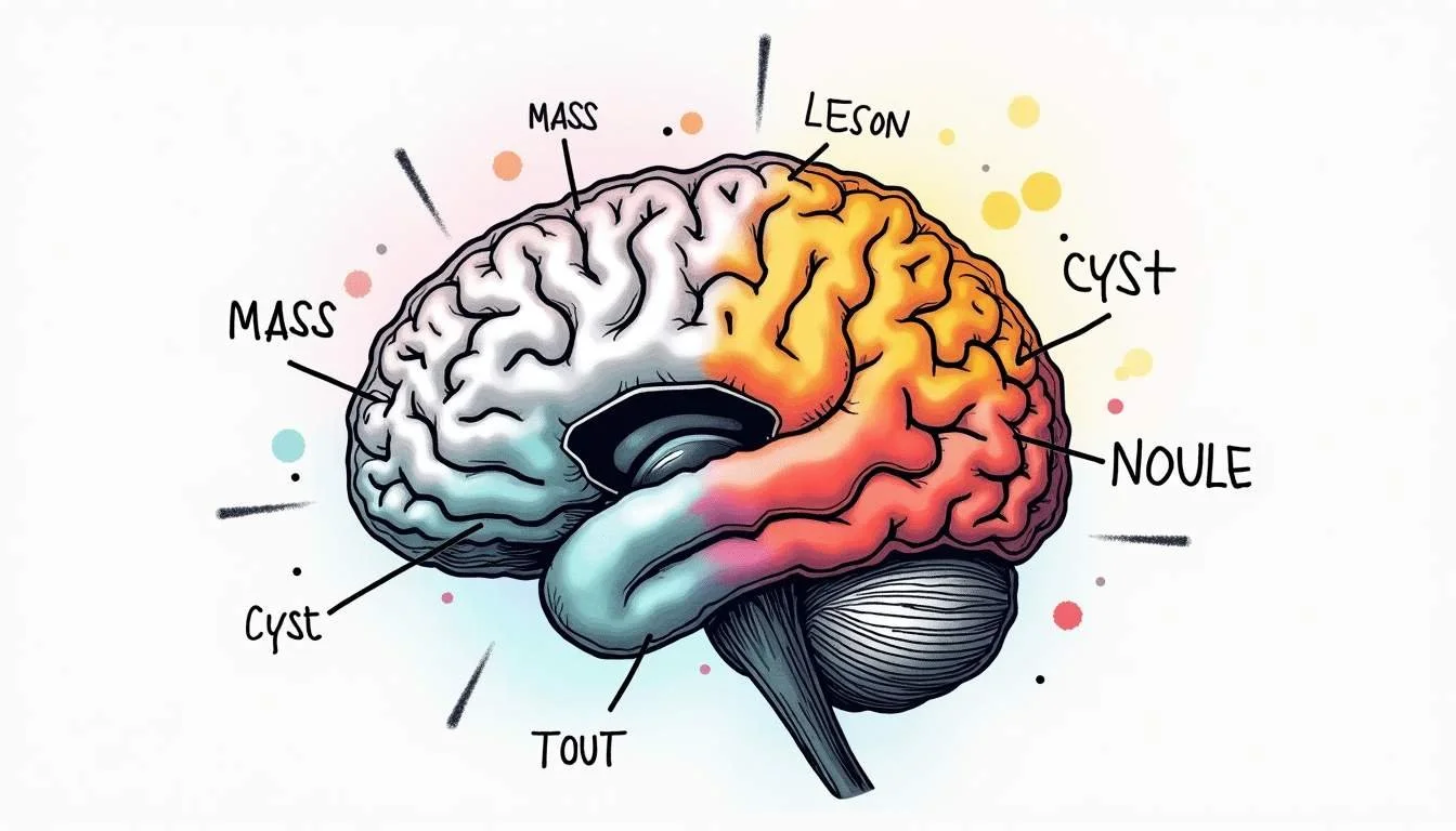
Mass, Nodule, Cyst: How They Differ
While “lesion” is the broad catch‑all, other terms such as “mass,” “nodule,” and “cyst” give extra clues about what the abnormal area looks like on the scan. These words still do not deliver a final diagnosis, but they hint at the structure and behavior of what the radiologist is seeing.
Think of these terms as describing the “shape and texture” of the finding, the same way a mechanic might say a part is solid, hollow, or fluid-filled before deciding what to do about it.
What “Mass” Usually Implies
“Mass” usually refers to a space‑occupying area that is larger or more substantial than the surrounding brain tissue. It tends to push, distort, or replace normal structures. A mass could be a tumor, but it could also be an abscess, a large cyst, or a localized collection of blood.
Because “mass” refers to how much space something takes up, it is often paired with other descriptors: solid, cystic, enhancing (after contrast), or infiltrative. Surgeons and radiologists sometimes use intraoperative tools such as ultrasound to refine what a mass is during surgery, distinguishing tumors from abscesses or hematomas based on how they look and feel in real time on imaging and pathology review of brain mass lesions as described in comparative studies of brain mass lesions.
What “Nodule” Suggests
“Nodule” usually describes a more rounded, often smaller focus that is distinct from surrounding tissue. On brain MRI, a nodule might show up inside another structure (for example within a cyst or gland) or on its own. Nodules can be benign or malignant; the term itself does not decide that. It just signals a relatively compact, focal spot that stands out on the images.
The report may describe how the nodule behaves with contrast, whether it has smooth or irregular borders, and what structures it touches. Those features help the treating team decide if it looks more like an infection, an inflammatory focus, a benign growth, or something that needs more urgent evaluation.
What Makes a “Cyst” Different
A “cyst” is an area that appears fluid‑filled on imaging. On MRI, fluid follows particular signal patterns, so radiologists can often tell when a lesion is mostly or entirely filled with liquid rather than solid tissue. Cysts can arise from normal developmental remnants, previous bleeding, infection, inflammation, or benign tumors that have a large fluid component.
Some cystic lesions in the brain are infectious or inflammatory and need prompt recognition, because the underlying process can respond to targeted treatment when caught early. Reviews of infectious and inflammatory cystic brain lesions stress that imaging patterns-how clear the borders are, how the surrounding brain reacts, and how the cyst behaves with contrast-are key clues that guide diagnosis and therapy choices in detailed neuroradiology analyses.
Unsure what your scan results really mean?
We analyze MRI, CT, PET, Ultrasound, and X-ray reports and deliver a clear, easy-to-understand summary in under 1 minute.
- Understand your results in simple language
- Easy to understand explanations
- Get a list of questions to ask your doctor
Incidental Findings on Brain MRI
Not every lesion or cyst discovered on a brain MRI is related to symptoms. Many are “incidental findings,” meaning they appear on a scan done for another reason, such as headaches, dizziness, or trauma. A common example is the pituitary incidentaloma-an unexpected lesion in the pituitary region found when the MRI was ordered for something else. Studies of brain MRI scans have reported that incidental pituitary lesions, often called pituitary incidentalomas, can be seen in a noticeable minority of patients, with published prevalence estimates ranging between 0.3% and 12.3% in some series in radiology research on incidental pituitary lesions.
For the person reading the report, “incidental” can sound dismissive when it actually means, “This might be unrelated to why the scan was done, but we still need to think through what it is and what to do.” The clinical question becomes: Does this finding pose any risk now or in the future? Does it need more testing, treatment, or just periodic monitoring?
A broad review of incidental findings on adult brain MRI emphasized that how these surprises are handled matters for both health and peace of mind. Appropriate management can prevent unnecessary anxiety and medical costs, while still catching the minority of incidental lesions that do need treatment or follow‑up imaging according to a comprehensive review of incidental MRI findings in adults. When something “incidental” shows up on a report, the next conversation with the ordering doctor is crucial for putting risk-and next steps-into perspective.
Special Case: Pituitary and Hypothalamic Lesions
Lesions in the sellar and parasellar region, home to the pituitary gland and nearby structures, deserve special mention because the words used here can sound especially worrying. MRI is the preferred imaging tool for this part of the brain, because it gives fine detail on tumors such as pituitary adenomas, craniopharyngiomas, and Rathke cleft cysts, along with their relationship to the optic nerves and surrounding vessels in dedicated imaging reviews of the hypothalamic–pituitary region.
Many pituitary lesions are benign and slow‑growing, and some are found in children and teenagers. A long‑term population‑based analysis looking at pediatric pituitary adenomas and cysts reported an overall incidence of 2.29 cases per 100,000 person‑years, with incidence rising in more recent decades in detailed epidemiologic work on pediatric pituitary lesions. Numbers like that highlight that these conditions are uncommon but not rare, and that more of them are being detected due to better and more frequent imaging.
Because the pituitary helps control hormones throughout the body, doctors often pair MRI findings with blood tests that look at hormone levels. A small “lesion” on a report may turn out to be a tiny, stable pituitary adenoma that only needs periodic checks. In other cases, symptoms such as vision changes or hormone imbalance guide decisions toward surgery or medical treatment.
How Doctors Work Out What a Lesion Really Is
Once a lesion, mass, cyst, or nodule is spotted on MRI, the medical team shifts from description to detective work. The radiologist’s report is the starting point: it lays out the size, location, signal characteristics, and how the lesion behaves with contrast. From there, the treating doctor weighs those details against the person’s symptoms, exam findings, and history.
For cystic or partially cystic lesions, subtle differences on imaging can point toward infection, inflammation, benign tumor, or something else. Narrative reviews of infectious and inflammatory cystic brain lesions describe how factors like the thickness of the cyst wall, the pattern of contrast enhancement, and the amount of surrounding brain swelling can help distinguish treatable infections from other mimics on MRI in specialized neuroradiology literature. Those distinctions matter because they change the urgency and type of treatment.
Sometimes imaging and clinical data still do not fully answer the question. In those cases, doctors may recommend follow‑up scans after a period of time, more advanced imaging sequences, functional tests (such as hormone assays for pituitary lesions), or, when appropriate, a biopsy or surgical removal. For many small, stable lesions that do not match worrisome patterns and are not causing symptoms, careful observation with periodic imaging is often the safest and most balanced choice.
Technology and the Future of Brain MRI Reports
Artificial intelligence is starting to assist radiologists in reading brain MRIs, especially for tumor detection and classification. Research using convolutional neural networks, a type of deep learning model, has reported very high accuracy when trained on MRI images of brain tumors, with one study from October demonstrating about 98% accuracy in distinguishing tumor types on imaging datasets in a detailed analysis of CNN performance on brain tumor MRI.
These tools are not a replacement for radiologists or clinical judgment, but they may help flag subtle lesions more consistently, suggest likely diagnoses, or prioritize scans that need urgent attention. Over time, that could mean more standardized descriptions of masses, nodules, cysts, and other lesions-and possibly clearer, more structured language that is easier to translate into patient‑friendly explanations.
How to Read Your Report and Talk With Your Doctor
When a brain MRI report lands in the patient portal, the temptation to dissect every word before talking with a doctor is strong. It can help to remember that terms like “lesion,” “mass,” “nodule,” and “cyst” start as descriptive labels, not final verdicts. The real interpretation happens when those words are combined with symptoms, exam findings, history, and, if needed, additional tests.
Reviews of incidental brain MRI findings stress that how these discoveries are communicated and managed can significantly influence both outcomes and anxiety levels in large‑scale analyses of adult MRI reports. Bringing questions to the follow‑up visit-What exactly does this term mean in my case? How confident are you in what this is? What are the realistic risks if we watch it?-turns a scary‑sounding report into a starting point for a clearer plan.
Mass, lesion, cyst, and nodule are technical words, but they do not have to stay mysterious. With a bit of translation and a chance to discuss the report in context, many people discover that the most frightening‑sounding phrases on the page point to something manageable, monitorable, or entirely benign.
Waiting for answers? You don’t have to.
Upload your MRI, CT, PET, Ultrasound, or X-ray report and receive a clear explanation in < 1 minute.
Take Control of Your MRI Understanding with Read My MRI
If the technical terms in your brain MRI report seem daunting, remember that clarity is just a click away. At Read My MRI, we're committed to demystifying your medical reports. Our AI-powered platform is designed to translate complex MRI, CT, PET, X-Ray, or Ultrasound reports into straightforward summaries. Say goodbye to medical jargon and hello to clear, concise explanations. Whether you're deciphering your own report or you're a healthcare provider seeking streamlined analysis, Get Your AI MRI Report Now! and experience the ease of understanding your medical imaging with Read My MRI.