What Does Calcification Mean in an MRI Report? Explained
When you receive an MRI report, it can often feel like reading a foreign language. Among the many technical terms, “calcification” is one that frequently appears and can cause concern. Understanding what calcification means in the context of an MRI report is essential for patients and caregivers alike. This article breaks down the concept of calcification, its significance in MRI findings, and what it might imply for your health.
Understanding Calcification: The Basics
Calcification refers to the accumulation of calcium salts in body tissues. While calcium is a vital mineral necessary for healthy bones and teeth, its presence in soft tissues or organs can sometimes indicate an abnormal process. This abnormal calcification can occur in various forms, including dystrophic calcification, which occurs in damaged or necrotic tissue, and metastatic calcification, which results from elevated calcium levels in the blood, often due to conditions such as hyperparathyroidism or cancer. Understanding the underlying causes of calcification is crucial for diagnosing and managing potential health issues.
In medical imaging, calcifications appear as dense, bright spots because calcium deposits block the passage of imaging signals differently than surrounding tissues. This makes them visible on various imaging modalities, including X-rays, CT scans, and sometimes MRI scans. The identification of these calcifications can provide critical insights into a patient’s health, as they may signify the presence of conditions such as atherosclerosis, tumors, or even chronic inflammation. Radiologists often categorize calcifications based on their morphology and distribution, which can help in determining their clinical significance.
However, it’s important to note that MRI is not the primary imaging technique for detecting calcifications. CT scans are generally more sensitive to calcium deposits due to their ability to capture differences in tissue density more clearly. Despite this, calcifications can still be noted on MRI, especially when they are large or in particular locations. In some cases, advanced MRI techniques, such as gradient echo sequences, can enhance the visibility of calcifications, allowing for a more comprehensive evaluation of the affected area. Understanding the imaging characteristics of calcifications can aid healthcare providers in making informed decisions regarding further diagnostic testing or treatment plans.
Additionally, the clinical implications of calcification can vary significantly based on its location and the patient's overall health. For instance, calcifications in the breast can raise concerns about breast cancer, prompting further investigation through biopsies or additional imaging. In contrast, calcifications in the kidneys may indicate chronic kidney disease or the presence of kidney stones, which can lead to different management strategies. As such, evaluating calcifications is not merely a matter of identifying their presence, but also involves a nuanced understanding of their potential impact on patient care and outcomes.
How Does Calcification Appear on an MRI?
Magnetic Resonance Imaging (MRI) uses magnetic fields and radio waves to create detailed images of the body’s internal structures. Unlike CT scans, MRI is excellent at visualizing soft tissues but less sensitive to calcium deposits.
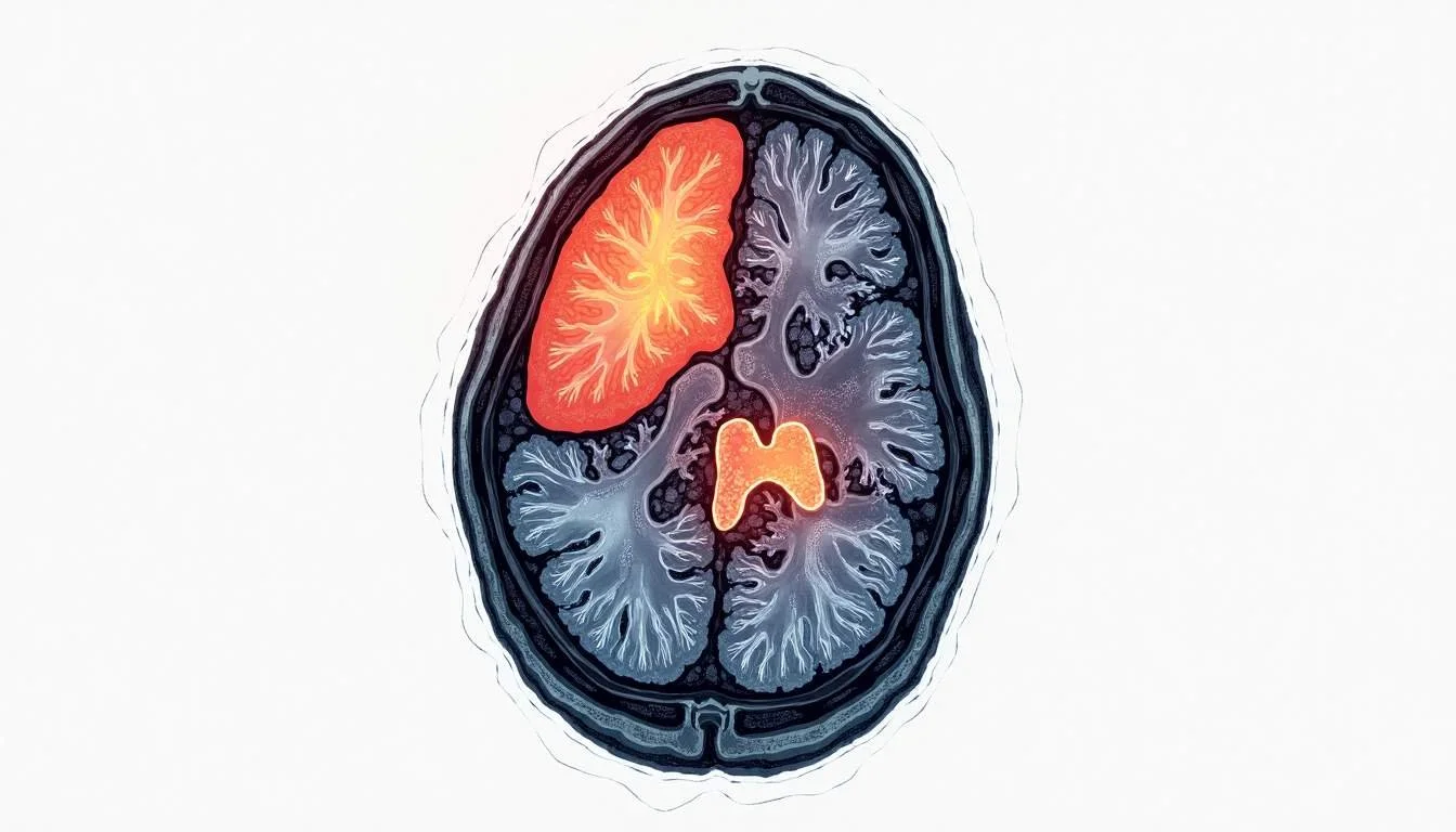
On MRI images, calcifications typically appear as areas of very low signal intensity, meaning they look dark or black on most MRI sequences. This is because calcium does not produce a signal in MRI, leading to a characteristic “signal void.”
However, distinguishing calcifications from other causes of low signal intensity, such as air pockets, metal implants, or certain types of hemorrhage, can be challenging. Radiologists use the location, shape, and context of these dark areas, alongside other imaging findings and clinical information, to interpret their significance. For instance, calcifications in the breast may indicate benign conditions like fibrocystic changes, while those in the brain could suggest more serious issues, such as tumors or vascular malformations.
Why MRI Is Less Sensitive to Calcifications
Calcium deposits are dense and lack free protons, which MRI relies upon to generate images. Since MRI detects signals from hydrogen protons in water and fat, calcifications, which contain little to no hydrogen, produce minimal signal. This results in dark spots on the images but does not always allow for precise characterization.
In contrast, CT scans measure tissue density directly, making calcium deposits stand out as bright white spots. This difference is why CT is often preferred when calcification is suspected or needs to be evaluated further. Additionally, the speed of CT imaging allows for rapid assessment in emergency situations, where time is critical, such as in cases of suspected stroke or acute trauma. Furthermore, the ability of CT to provide a clear depiction of bony structures and calcifications makes it invaluable in orthopedic evaluations, where understanding the extent of calcification can influence treatment decisions.
Common Causes of Calcification Seen on MRI
Calcifications can occur in various tissues and organs for many reasons. Some calcifications are harmless and part of the normal aging process, while others may signal underlying disease.
1. Vascular Calcification
One of the most frequent types of calcification seen on imaging involves blood vessels. Vascular calcification happens when calcium builds up in the walls of arteries, often as a result of atherosclerosis (hardening of the arteries).
This process is common in older adults and those with risk factors such as diabetes, high cholesterol, or hypertension. While vascular calcification can stiffen arteries and contribute to cardiovascular disease, small amounts are often considered a normal part of the aging process. Interestingly, the presence of vascular calcification can also serve as a marker for cardiovascular risk, prompting healthcare providers to implement preventive measures such as lifestyle modifications or medications aimed at reducing cholesterol levels and improving vascular health.
2. Brain Calcifications
Calcifications in the brain can be detected on MRI, particularly when they are large or located in specific areas, such as the basal ganglia, pineal gland, or choroid plexus. Some brain calcifications are benign and incidental findings, while others may be associated with infections, metabolic disorders, or tumors.
For example, certain infections such as toxoplasmosis or cytomegalovirus can cause calcifications in brain tissue. Additionally, certain genetic conditions can lead to abnormal calcium deposits in the brain, potentially affecting neurological function. In particular, conditions like Fahr's syndrome, characterized by bilateral calcifications in the basal ganglia, can lead to a range of neurological symptoms, including movement disorders and cognitive decline, highlighting the importance of identifying the underlying cause of such calcifications.
3. Tumor-Related Calcifications
Some tumors develop calcifications within their mass. These calcifications can help radiologists differentiate between types of tumors and distinguish between benign and malignant growths. For instance, calcifications are often seen in meningiomas (a type of brain tumor) and certain breast tumors.
In these cases, the pattern and extent of calcification provide clues about the tumor’s nature and behavior, aiding diagnosis and treatment planning. The presence of microcalcifications in breast tissue, for example, can be indicative of ductal carcinoma in situ (DCIS), a non-invasive form of breast cancer. Radiologists often use the morphology of these calcifications—such as their shape, size, and distribution—to assess the likelihood of malignancy and guide further diagnostic procedures like biopsies.
4. Soft Tissue and Organ Calcifications
Calcifications can also form in soft tissues and organs such as the lungs, kidneys, and breasts. For example, in the lungs, calcifications may result from prior infections, such as tuberculosis or histoplasmosis. In the kidneys, calcifications may indicate kidney stones or chronic inflammation.
In the breast, calcifications are commonly detected on mammograms and can be benign or suggestive of early cancer, depending on their appearance. Moreover, in the kidneys, the presence of calcifications can also be associated with conditions such as renal artery stenosis or chronic kidney disease, where the calcium deposits may reflect underlying vascular issues or metabolic imbalances. Understanding the context of these calcifications is crucial for healthcare providers, as it can influence management strategies and the need for further investigation or intervention.
Interpreting Calcification in Your MRI Report
When your MRI report mentions calcification, it is important to understand the context in which it appears. Calcifications themselves are not diseases, but instead signs that may indicate underlying conditions.
Radiologists typically describe calcifications in terms of their size, shape, location, and number. These details help your healthcare provider determine whether further evaluation or treatment is necessary.
Benign vs. Concerning Calcifications
Many calcifications are benign and require no treatment. For example, small vascular calcifications or incidental brain calcifications in older adults often have no clinical significance.
However, certain patterns of calcification may warrant additional investigation. For example, irregular or clustered calcifications in a tumor might suggest malignancy, prompting biopsy or further imaging.
Follow-Up and Additional Testing
If calcifications are detected on an MRI and their significance is unclear, your doctor may recommend additional imaging, such as a CT scan, which provides a more detailed characterization of calcium deposits. Blood tests or biopsies might also be necessary, depending on the suspected cause.
It’s essential to discuss your MRI findings with your healthcare provider to understand the significance of the calcifications in your specific situation and determine whether any action is necessary.
Common Questions About Calcification in MRI Reports
Does Calcification Mean Cancer?
Not necessarily. Calcification can be present in both benign and malignant conditions. While some cancers may contain calcifications, many benign processes also cause calcium deposits. The pattern and context of calcification, along with other clinical information, guide the diagnosis.
Can Calcifications Cause Symptoms?
Calcifications themselves often do not cause symptoms. However, if they are part of a disease process, such as a tumor or vascular disease, symptoms may arise from the underlying condition rather than the calcification directly.
Is Calcification Reversible?
Calcification is generally considered a permanent deposit of calcium in tissues. While some medical treatments may slow progression or address underlying causes, existing calcifications typically do not disappear.
What to Take Away About Calcification in MRI Reports
Calcification in an MRI report is a descriptive term indicating the presence of calcium deposits within tissues. Although MRI is not the most sensitive tool for detecting calcifications, these findings can provide valuable clues about your health.
Understanding the type, location, and pattern of calcifications helps healthcare providers determine whether they are harmless or require further evaluation. If you encounter this term in your MRI report, it’s important to follow up with your doctor to interpret the findings in the context of your overall health and symptoms.
Ultimately, calcifications are common and often benign; however, they can also be markers of underlying conditions that may require attention. Staying informed and proactive about your imaging results is key to effective healthcare management.
Take Control of Your Health with Read My MRI
If you've encountered the term 'calcification' in your MRI report and are seeking a clearer understanding of what it means for your health, Read My MRI is here to help. Our AI-powered platform simplifies complex medical reports, converting them into easy-to-read summaries that are free from confusing medical jargon. Whether you're navigating a recent diagnosis or are a healthcare professional in need of streamlined report analysis, our service is designed to make medical information accessible and understandable. Don't let uncertainty about your MRI findings cause you stress. Get Your AI MRI Report Now! and gain the clarity you need to make informed decisions about your health care.

